Visit 1 · exam
3D CT Bone Assessment
3D CT assesses the height, thickness and density of the jawbone. The clinician combines the scan with the examination to plan where and how much bone augmentation may be needed.

Implant Center · Gangnam · Cheongdam · Bucheon
When jawbone height or thickness is insufficient, grafting can rebuild bone to support an implant. The graft and implant may be placed together or in separate stages after assessment. The clinician uses 3D CT and the clinical findings to plan materials and treatment; results vary.
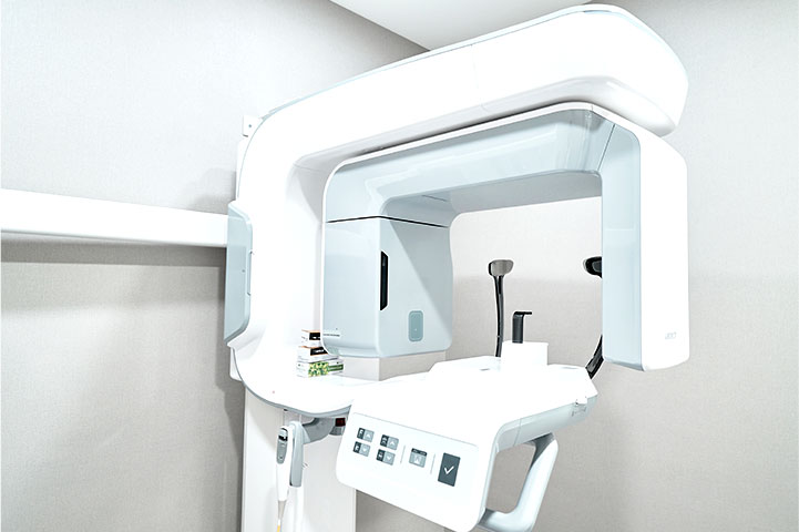
Visit 1 · exam
3D CT assesses the height, thickness and density of the jawbone. The clinician combines the scan with the examination to plan where and how much bone augmentation may be needed.
Visit 1 · consultation
A specialist experienced in bone grafting reviews your scan with you and customizes the graft material and surgical plan to your bone condition and overall health.
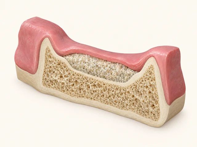
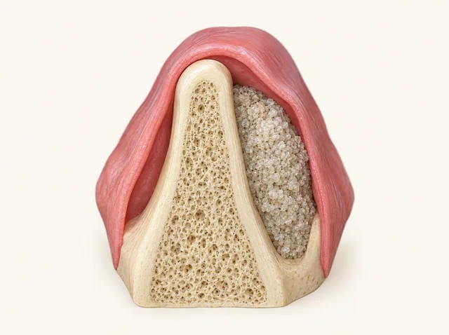
Find the closest situation. The clinician assesses your examination, 3D CT and overall health; this page cannot diagnose your case.
If gum disease has caused bone loss, the scan helps show how much bone remains and where. The clinician combines it with the oral examination and other clinical findings to plan whether grafting is needed.
After a tooth has been missing for a long time, the jawbone may have shrunk. The remaining height and thickness are part of the clinical assessment of whether grafting and implant placement can be simultaneous or need separate stages.
If the ridge is too thin or short to support the planned implant, a graft may be considered. Materials are selected for the defect, and the clinician assesses whether grafting and placement can be simultaneous or need separate stages.
If the ridge has changed after long-term denture use, a specialist reviews the scan and clinical findings with you to plan any grafting needed for your bone condition.
This page is not a diagnosis. The method and stages are planned after an oral examination and 3D CT, and may be revised according to clinical findings.
Assessment determines whether grafting and implant placement can be done together or need separate stages.
The graft is placed to rebuild the bone foundation. Depending on the condition of the site, the implant may be placed in the same procedure or in a later stage after the graft has matured.
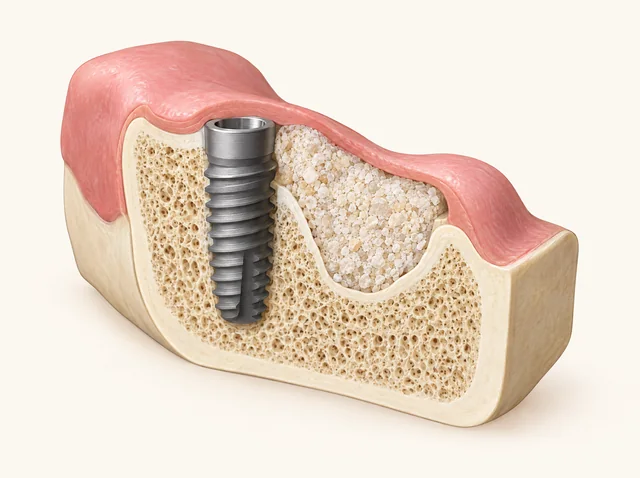
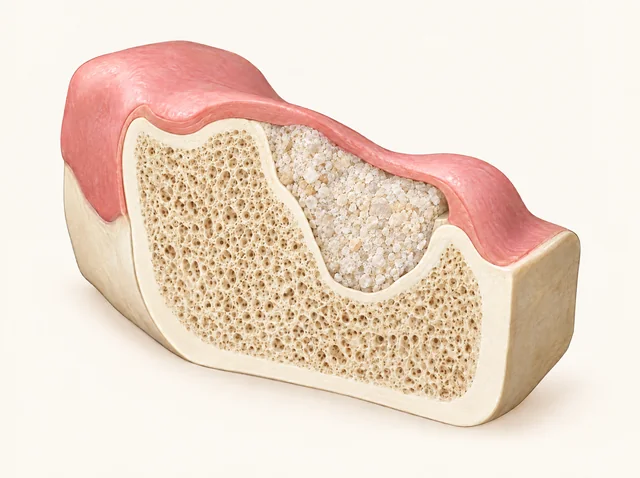
Bone height, thickness and density on 3D CT are part of the assessment. The clinician also considers the site and overall health when planning simultaneous or staged placement.
Healing speed varies. The initial visit plan follows diagnosis and is reviewed as healing progresses before later travel is arranged.
The clinician selects materials after assessing the defect size and location, bone quality, oral examination and 3D CT. The proposed material is discussed before treatment.
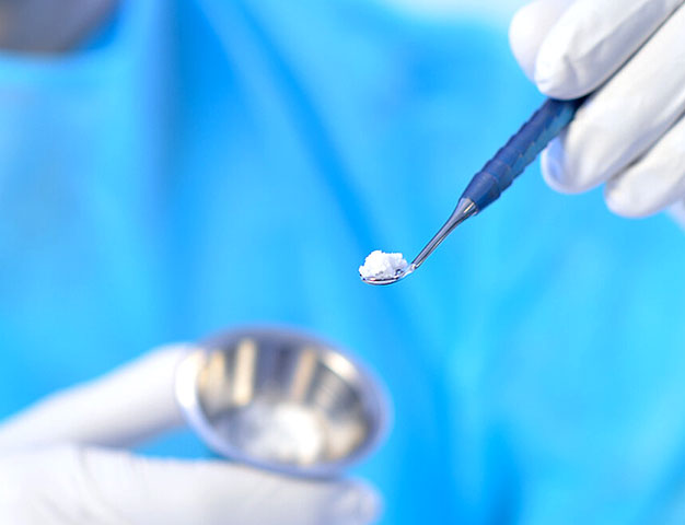
In suitable cases, an extracted tooth can be processed into graft material. The clinician assesses whether this option suits your case and discusses the proposed material before treatment.
The fixture, abutment and crown have different roles. A graft rebuilds supporting bone, either with implant placement or in an earlier stage according to the treatment plan.
The fixture is held by the bone around it. When the ridge lacks height or thickness, the graft rebuilds that bone before or during placement.
The abutment connects the fixture and the crown through the gum. It goes on once the implant is stable in the rebuilt bone.
The crown is connected after the implant integrates with the rebuilt bone, and the result is verified with precise imaging and bite checks.
After integration
After the implant has integrated with the rebuilt bone, the prosthesis is connected and checked with imaging and a bite review.
Long term
Periodic check-ups and maintenance guidance help care for the grafted bone and implant over the long term.
An ISQ reading of how firmly the fixture is held by bone.
The clinician considers stability readings together with healing and other clinical findings to plan the prosthesis; a reading alone does not set the date.
The finished result is photographed and the bite is checked.
Imaging and bite checks review the prosthesis after it is connected to the implant.
After the graft
Depending on your condition, the implant is placed with the graft or in a separate stage after the bone matures.
Healing speed varies by patient. The timeline is planned after diagnosis and reviewed during healing rather than guaranteed in advance.
Before the final tooth
Once the implant integrates with the rebuilt bone, the prosthesis is connected.
The treatment result is verified with precise imaging and bite checks.
Long term
Periodic check-ups and maintenance guidance help the grafted bone and implant stay healthy over the long term.
Reminder notices are sent for your check-up schedule.
The clinician selects graft materials according to the defect size and location, bone quality and clinical assessment, taking biocompatibility into account. Material selection does not remove treatment risks.
A graft can need more healing than planned, and an implant can fail to integrate. That is why stability is measured before the prosthesis is connected. Individual results vary.
Up to 5 years. Check-up guidance and a warranty certificate. Conditions apply.
Panoramic X-rays recorded at the clinic, dated on the film.
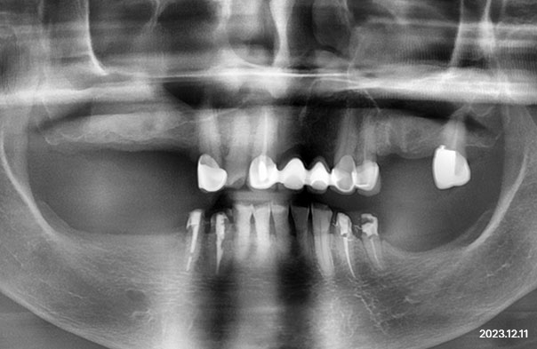
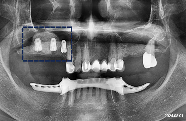
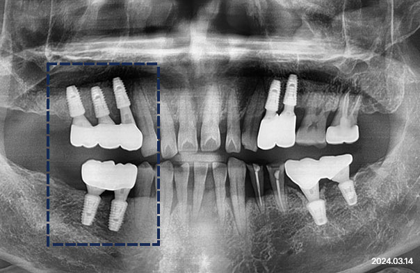
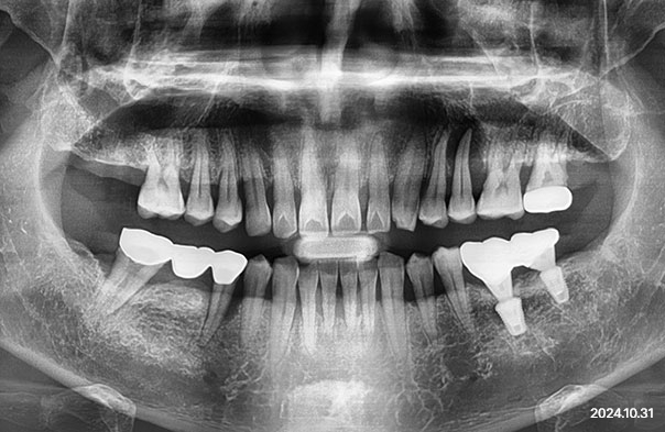
Records taken at the clinic, published as provided. Results vary by person, and treatment carries risks and possible side effects - see the recovery and precautions section on this page. Published with the patient's written consent for promotional use; dates as recorded.
A plan that touches gums, bone and restoration is discussed by the specialists in each field first.

Periodontics
Board-certified specialist in Periodontics (Ministry of Health and Welfare, Korea)
View full profile : Dr. Son Yeo-jin
Integrated Dentistry
Board-certified specialist in Integrated Dentistry (Ministry of Health and Welfare, Korea)
View full profile : Dr. Yoon So-jeongAn implant needs sufficient healthy bone for support. If bone is too thin or short, the implant may loosen or fail. Grafting aims to rebuild support and may be performed with placement or as an earlier stage after assessment. It does not guarantee long-term success, and individual results vary.
The clinician selects materials after assessing the defect size and location, bone quality, oral examination and 3D CT. Options include autogenous bone, allograft, xenograft and synthetic bone. In suitable cases, material made from your own extracted tooth may be considered. The proposed material is discussed before treatment.
In suitable cases, grafting and placement can share one surgery. If the graft needs to heal first, the implant is placed later. Healing speed varies, and bone integration, the final prosthesis and follow-up visits still need planning. Travel is discussed after clinical assessment; a short stay does not determine the treatment sequence.
Periodic check-ups and maintenance guidance, with reminder notices for your check-up schedule.
In suitable cases, an extracted tooth can be processed into graft material. The clinician assesses whether this option suits your case and discusses the proposed material before treatment.